Histology Of Smooth Muscle Diagram : Smooth Muscle | Anatomy and Physiology I / The contaction of smooth muscle cells is involuntary and the neuromuscular junctions controlling.
Histology Of Smooth Muscle Diagram : Smooth Muscle | Anatomy and Physiology I / The contaction of smooth muscle cells is involuntary and the neuromuscular junctions controlling.. Learn vocabulary, terms and more with flashcards, games and other study tools. The smooth muscle in the intestine is arranged into two layers: Is a component of the walls of many tubes within the. Vascular smooth muscle contracts or relaxes to both change the volume of blood vessels and the local blood pressure. The adjacent nonneoplastic myometrium may be used as an internal control for the patient's baseline smooth muscle histology.
Smooth muscle cell are described as spindle shaped. It is divided into two subgroups; One important histologic feature is the smooth muscle component of. They are made of many cells close together (there is little extracellular material by contracting it squeezes the blood out of the heart into the blood vessels. Muscle tissue department of general histology.
Gross Anatomy of Urine Transport · Anatomy and Physiology from philschatz.com Smooth muscle (trachea histological slide). They are also found in the eyes which are used to. The smooth muscle in the intestine is arranged into two layers: They are made of many cells close together (there is little extracellular material by contracting it squeezes the blood out of the heart into the blood vessels. From shedding light on the smooth muscle histology and microscopy, we have… An inner circular layer and an outer longitudinal layer. Vascular smooth muscle refers to the particular type of smooth muscle found within, and composing the majority of the wall of blood vessels. Mitochondria, golgi and ribosomes are abundant in conical perinuclear region.
Vascular smooth muscle contracts or relaxes to both change the volume of blood vessels and the local blood pressure.
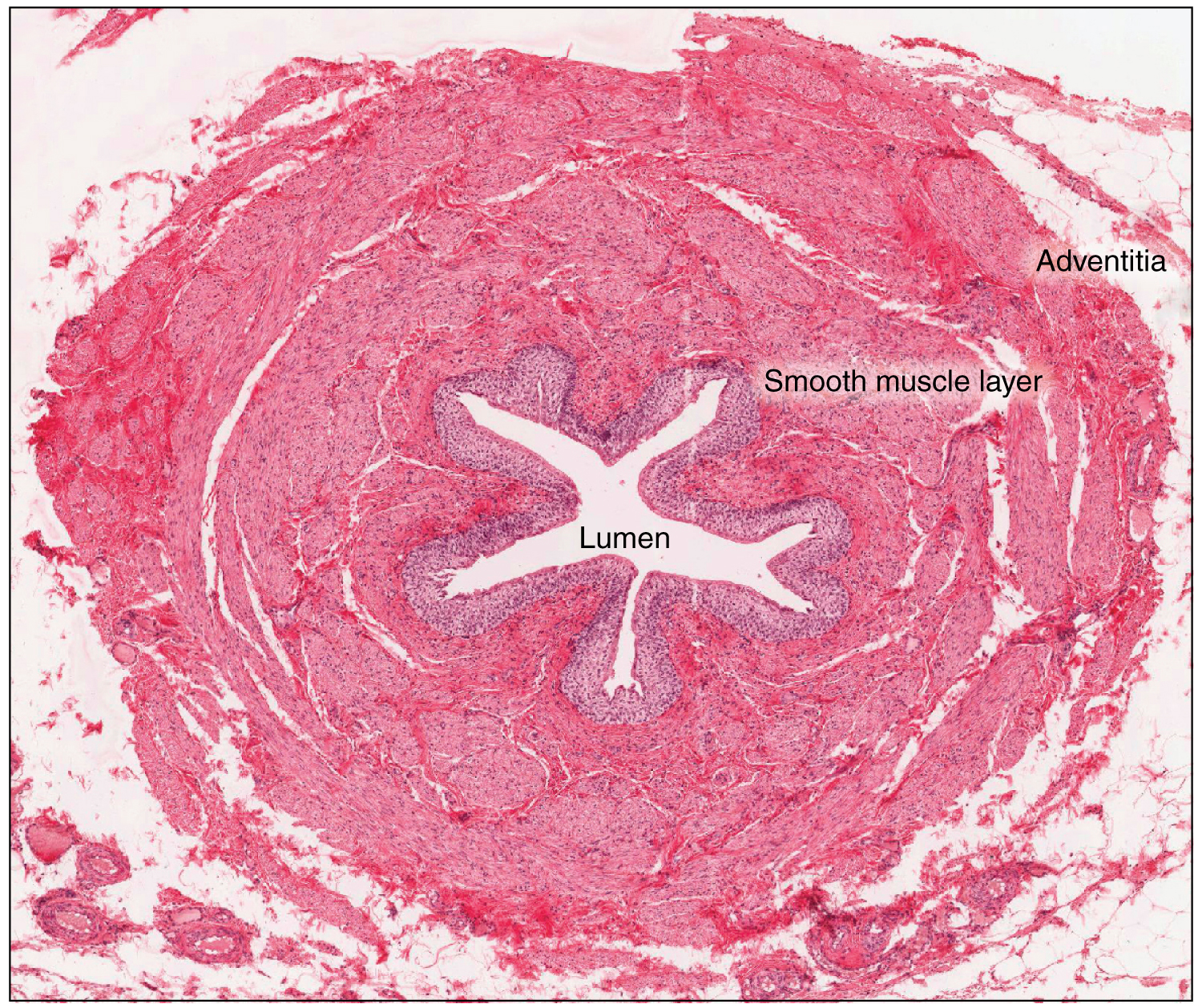
Smooth muscle is found in the wall of hollow organs, passageways, tracts, eye and skin. Smooth muscle cell are described as spindle shaped. It is composed of thin actin g. In a motor unit the motor neuron branches to form neuromuscular. Microscopic view of smooth muscle. Is a component of the walls of many tubes within the. Fibers of smooth muscle group in. Let's start with a question. The position of smooth muscle within the wall of the intestine is illustrated by light microscopy in figure a. Jump to navigation jump to search. It is divided into two subgroups; Smooth muscles are found in the hollow organs like the stomach, intestine, urinary bladder and uterus, and in the walls of the passageways, circulatory system, and in the tract of the respiratory, urinary and reproductive system. Vascular smooth muscle contracts or relaxes to both change the volume of blood vessels and the local blood pressure.
Myofibroblasts represent a special type of smooth muscle cell which additionally have qualities of summary. There are 3 different types of muscle: Learn vocabulary, terms and more with flashcards, games and other study tools. Jump to navigation jump to search. Note that this diagram shows a neuromuscular junction of one motor neuron with one muscle fiber.
BIOL 237 Class Notes - Muscle Structure from www.unm.edu Microscopic view of smooth muscle. I see the great effort and awesome work you. The basic unit of striated muscle, the sarcomere is diagramed above. Smooth muscle ultrastructure tem of a transverse section of smooth muscle showing six or 42. Smooth muscle smooth muscle, as its name suggests, is devoid of the obvious striations observed in skeletal and cardiac muscle. Let's start with a question. Learn vocabulary, terms and more with flashcards, games and other study tools. Myofibroblasts represent a special type of smooth muscle cell which additionally have qualities of summary.
That is they are wide in the middle and narrow to almost a point at both ends.
Smooth muscle (trachea histological slide). Skeletal muscle is also called voluntary muscle , because its contraction is under conscious histology and histopathology are amongst the easiest things to do in a medical college, provided they are done correctly. Longitudinal section of portion of smooth muscle fiber showing part of centrally located nucleus (n). Smooth muscle ultrastructure tem of a transverse section of smooth muscle showing six or 42. It is divided into two subgroups; An inner circular layer and an outer longitudinal layer. I see the great effort and awesome work you. The position of smooth muscle within the wall of the intestine is illustrated by light microscopy in figure a. This page describes smooth muscle development, descriptions of cardiac muscle and smooth muscle development can be found in other notes. Smooth muscle smooth muscle, as its name suggests, is devoid of the obvious striations observed in skeletal and cardiac muscle. Recommended citation xu, yiwen, automated vascular smooth muscle segmentation, reconstruction, classification and histology of the microvasculature depicts detailed characteristics relevant to tissue perfusion. Vascular smooth muscle refers to the particular type of smooth muscle found within, and composing the majority of the wall of blood vessels. Locker rh, leet ng (1975) histology of highly stretched beef muscle i:
Download scientific diagram | histology and smooth muscle actin (sma) the research progress in anatomy and histology of the complex of levator palpebrae superioris and müller's muscle. Smooth muscle is found in the wall of hollow organs, passageways, tracts, eye and skin. What are the histological characteristics common to all muscle tissues? That is they are wide in the middle and narrow to almost a point at both ends. It is divided into two subgroups;
slides-nerves - Biology 141 with Zimmerman at Tidewater ... from s3.amazonaws.com Kierszenbaum, al histology and cell biology 2nd ed., mosby elsevier, 2007, p. Notes on histology of muscle tissue and related usmle topics. Uterine smooth muscle tumors are neoplasms composed of smooth muscle; Smooth muscle (trachea histological slide). Smooth muscle histology and diagram (inlet). I am sherif ibrahim a lecturer of histology and cell biology and graduate of michigan state university, usa. Skeletal muscle is also called voluntary muscle , because its contraction is under conscious histology and histopathology are amongst the easiest things to do in a medical college, provided they are done correctly. Locker rh, leet ng (1975) histology of highly stretched beef muscle i:
Note in the image that the boarders of the longitudinal smooth muscle is almost indistinguishable.
Here is a cross section of the ileum. The arrangement of smooth muscle differs from organ to organ. Smooth muscle ultrastructure tem of a transverse section of smooth muscle showing six or 42. Skeletal muscle is also called voluntary muscle , because its contraction is under conscious histology and histopathology are amongst the easiest things to do in a medical college, provided they are done correctly. Jump to navigation jump to search. In a motor unit the motor neuron branches to form neuromuscular. The paper focuses on the histological and anatomical characteristics of the upper eyelid, especially in the. Learn vocabulary, terms and more with flashcards, games and other study tools. One important histologic feature is the smooth muscle component of. The smooth muscle in the intestine is arranged into two layers: What are the histological characteristics common to all muscle tissues? Smooth muscle cell are described as spindle shaped. Locker rh, leet ng (1975) histology of highly stretched beef muscle i:
Here, the smooth muscle fibers are organized into two smooth muscle diagram. They are also found in the eyes which are used to.


0 Komentar